15/10/02说明:此前论坛服务器频繁出错,现已更换服务器。今后论坛继续数据库备份,不备份上传附件。

肝胆相照论坛


肝胆相照论坛 论坛 学术讨论& HBV English 完整和不完整的乙型肝炎病毒颗粒:形成,功能和应用 ...
查看: 1282|回复: 10
go

完整和不完整的乙型肝炎病毒颗粒:形成,功能和应用 [复制链接]

Rank: 8Rank: 8

现金
62111 元 
精华
26 
帖子
30441 
注册时间
2009-10-5 
最后登录
2022-12-28 

才高八斗

1
发表于 2017-3-29 20:54 |只看该作者 |倒序浏览 |打印
Viruses 2017, 9(3), 56; doi:10.3390/v9030056
                                Complete and Incomplete Hepatitis B Virus Particles: Formation, Function, and Application                Jianming Hu 1,*                                                                                                                      and Kuancheng Liu 1,2
               
                                                                                                                        1
                                        Department of Microbiology and Immunology, Penn State University College of Medicine, Hershey, PA 17033, USA
               
                                                                                2
                                        College of Life Sciences, Zhejiang Sci-Tech University, Hangzhou 310018, China
               
                                                               
                                                                        *
                Author to whom correspondence should be addressed.
            
               
                        Academic Editors: Ulrike Protzer and Michael Nassal
                            Received: 8 February 2017 / Revised: 11 March 2017 / Accepted: 17 March 2017 / Published: 21 March 2017        
                                            (This article belongs to the  Special Issue Recent Advances in Hepatitis B Virus Research)            
                                                                                                                                        View Full-Text                                                                                       |                                                                                                      Download PDF [576 KB, uploaded 21 March 2017]                                                                                                                              |                                                                           [url=]Browse Figures[/url]                        


                                                                                                                                                                                                                                                Abstract Hepatitis B virus (HBV) is a para-retrovirus or retroid virus that contains a double-stranded DNA genome and replicates this DNA via reverse transcription of a RNA pregenome. Viral reverse transcription takes place within a capsid upon packaging of the RNA and the viral reverse transcriptase. A major characteristic of HBV replication is the selection of capsids containing the double-stranded DNA, but not those containing the RNA or the single-stranded DNA replication intermediate, for envelopment during virion secretion. The complete HBV virion particles thus contain an outer envelope, studded with viral envelope proteins, that encloses the capsid, which, in turn, encapsidates the double-stranded DNA genome. Furthermore, HBV morphogenesis is characterized by the release of subviral particles that are several orders of magnitude more abundant than the complete virions. One class of subviral particles are the classical surface antigen particles (Australian antigen) that contain only the viral envelope proteins, whereas the more recently discovered genome-free (empty) virions contain both the envelope and capsid but no genome. In addition, recent evidence suggests that low levels of RNA-containing particles may be released, after all. We will summarize what is currently known about how the complete and incomplete HBV particles are assembled. We will discuss briefly the functions of the subviral particles, which remain largely unknown. Finally, we will explore the utility of the subviral particles, particularly, the potential of empty virions and putative RNA virions as diagnostic markers and the potential of empty virons as a vaccine candidate.                                                                                                                                View Full-Text                                        
                                                    Keywords:                                                                                                                                                                               hepatitis B virus;                                virion;                                empty virion;                                Australian antigen;                                HBsAg;                                HBcAg;                                subviral particles;                                CCC DNA;                                diagnosis;                                vaccine                                    
                                                                                                                                                                                ▼            Figures


Figure 1


                                                                                                            This is an open access article distributed under the Creative Commons Attribution License which permits unrestricted use, distribution, and reproduction in any medium, provided the original work is properly cited. (CC BY 4.0).                                       
                  

Rank: 8Rank: 8

现金
62111 元 
精华
26 
帖子
30441 
注册时间
2009-10-5 
最后登录
2022-12-28 

才高八斗

2
发表于 2017-3-29 20:54 |只看该作者
病毒2017,9(3),56; doi:10.3390 / v9030056
完整和不完整的乙型肝炎病毒颗粒:形成,功能和应用
刘建明1,*,刘冠成1,2
1
美国宾夕法尼亚州立大学医学院微生物与免疫学系,美国宾夕法尼亚州赫尔案17033号
2
浙江科技大学生命科学学院,杭州310018
*
作者应向谁传递信函。
学术编辑:Ulrike Protzer和Michael Nassal
收到日期:2017年2月8日/修订日期:2017年3月11日/接受日期:2017年3月17日/发布日期:2017年3月21日
(本文属于乙肝病毒研究最新进展的特刊)
查看全文|下载PDF [576 KB,于2017年3月21日上载]浏览数字
抽象
乙型肝炎病毒(HBV)是一种副反转录病毒或网状病毒,含有双链DNA基因组,并通过RNA pregenome的逆转录复制该DNA。病毒逆转录在RNA和病毒逆转录酶包装后在衣壳内进行。 HBV复制的主要特征是选择含有双链DNA的衣壳,但不包含含有RNA或单链DNA复制中间体的衣壳,用于在病毒体分泌过程中包膜。因此,完整的HBV病毒粒子包含外壳,其上散布有病毒包膜蛋白,其包围衣壳,其反过来包裹双链DNA基因组。此外,HBV形态发生的特征在于释放比完整病毒体更多数量级的亚病毒颗粒。一类亚病毒颗粒是仅含有病毒包膜蛋白的经典表面抗原颗粒(澳大利亚抗原),而最近发现的无基因组(空)病毒粒子包含包膜和衣壳,但不含基因组。此外,最近的证据表明,低含量的含RNA颗粒可能会被释放。我们将总结目前已知的完整和不完整的HBV颗粒如何组装。我们将简要讨论亚病毒颗粒的功能,这些功能在很大程度上是未知的。最后,我们将探讨亚病毒颗粒的用途,特别是潜在的空载体病毒粒子和推定的RNA病毒粒子作为诊断标记物,以及潜在的空白病毒作为候选疫苗。查看全文
关键词:乙型肝炎病毒;病毒粒子空病毒粒子;澳大利亚抗原HBsAg; HBcAg;亚病毒颗粒CCC DNA;诊断;疫苗
▼数字

图1
这是一种根据知识共享署名许可方式分发的开放获取文章,允许在任何媒体中不受限制地使用,分发和复制,前提是原始作品被正确引用。 (CC BY 4.0)。

Rank: 8Rank: 8

现金
62111 元 
精华
26 
帖子
30441 
注册时间
2009-10-5 
最后登录
2022-12-28 

才高八斗

3
发表于 2017-3-29 21:04 |只看该作者
Do Empty Virions Contain an Aberrant Core Protein, HBcrAg?
Over 10 years ago, there was a report claiming that an aberrant protein derived from the aforementioned PreCore protein, dubbed HBc related antigen or HBcrAg, in some DNA-free HBV virions in patient sera [47]. The protein migrated as a 22 kd protein (hence p22cr) on sodium dodecyl sulfate-acrylamide gels. Western blot analysis using HBc/HBe specific antibodies and mass spectrometry appeared to identify HBcrAg as an aberrantly processed PreCore protein containing the entire PreC region including the uncleaved signal peptide (i.e., with a 29 residue N-terminal extension relative to HBc or 19 residue extension relative to HBe) but similar to HBe, lacking the C-terminal arginine-rich domain or CTD (ending at ca. 150), which mediates RNA and DNA binding by HBc. It remains unclear how this aberrant PreCore protein could assemble into capsids as the PreC region is known to disrupt capsid assembly via an intramolecular disulfide bond involving a Cys residue in the PreC region [14], or if the presumed capsid is made, how it would be enveloped and secreted. No follow-up studies have been reported by the authors or others to confirm this report. On the other hand, we have shown that empty virions clearly contain capsids consisting of the normal HBc protein and no aberrant core related protein or any PreC region sequence is needed for empty virion secretion [8]. Furthermore, using multiple antibodies specific for the HBc CTD, which was supposedly missing from HBcrAg in empty virions, including one antibody whose epitope includes the last Cys (183) residue [49], we could detect the normal HBc protein in empty virions in cell culture supernatant and in human serum samples, indicating clearly they contain the entire CTD [43]. At this point, it seems likely that the so-called HBcrAg might have been an artifact of detection and in any case, it is certainly not required for empty virion secretion.

做空的病毒含有异常核心蛋白,HBcrAg?
10多年前,有报道称,在患者血清中的一些无DNA的HBV病毒粒子中,衍生自上述PreCore蛋白质的异常蛋白质被称为HBc相关抗原或HBcrAg [47]。该蛋白质在十二烷基硫酸盐 - 丙烯酰胺凝胶上以22kd蛋白质(因此p22cr)迁移。使用HBc / HBe特异性抗体和质谱的蛋白质印迹分析似乎将HBcrAg鉴定为含有包含未切割信号肽的整个PreC区域的异常加工的PreCore蛋白(即,具有相对于HBc或19残基延伸的29个残基的N末端延伸相对于HBe),但类似于HBe,缺乏C末端富含精氨酸的结构域或CTD(终止于约150),其介导HBc的RNA和DNA结合。目前还不清楚这种异常的PreCore蛋白如何组装成衣壳,因为已知PreC区域通过涉及PreC区域[C]中的Cys残基的分子内二硫键破坏衣壳组装,或者如果假定的衣壳是如何被包围和分泌。作者或其他人没有报告后续研究来确认本报告。另一方面,我们已经显示空的病毒粒子清楚地包含由正常的HBc蛋白组成的衣壳,没有异常的核心相关蛋白质,或者空的病毒体分泌物需要任何PreC区域序列[8]。此外,使用对于空白病毒体中HBcrAg缺失的HBc CTD特异的多种抗体,包括一个抗体,其表位包括最后一个Cys(183)残基[49],我们可以检测细胞中空的病毒粒子中的正常HBc蛋白培养上清液和人血清样品,表明它们含有整个CTD [43]。在这一点上,所谓的HBcrAg似乎可能是检测的工具,在任何情况下,绝对不需要空的病毒体分泌。

Rank: 8Rank: 8

现金
62111 元 
精华
26 
帖子
30441 
注册时间
2009-10-5 
最后登录
2022-12-28 

才高八斗

4
发表于 2017-3-29 21:05 |只看该作者
2.4. RNA Virions
Even more surprising than the discovery of empty HBV virions, it appears that HBV may also secrete, albeit at a low level, RNA-containing particles (Figure 1), thus blurring the distinction between para-retroviruses and conventional retroviruses. Apparently inspired by the need to identify easily accessible surrogate markers for monitoring hepatic CCC DNA, there have been a flurry of reports recently on the detection of HBV RNA in serum samples of HBV infected patients [10,11,12,13,57,58]. Most of these suggest that HBV RNA levels detected in the patient sera are around 106 copies/mL, ca. 0.1%–1% of HBV DNA levels, in the absence of antiviral treatment [10,11,13,57,59,60].
The nature of the viral RNA as well as the physical entity that the RNA is associated with in the blood remain to be better characterized. Some evidence has been reported suggesting that the HBV RNA in serum may be associated with viral capsids as demonstrated by anti-HBc immunoprecipitation [57]. The fact that detergent treatment enhanced the immunoprecipitation efficiency was consistent with a possible association of the RNA with enveloped virions [57], although other membranous vesicles were not excluded. It remains possible that at least some HBV RNA detected in the serum samples is released in vesicles, e.g., as a result of lysis of infected hepatocytes, independent of viral capsid or virion formation. Consistent with this possibility is the detection of truncated HBV RNAs, perhaps as fusion to cellular RNAs and transcribed from integrated HBV DNA, were detected in the blood, in the absence of any other HBV markers [11,58].
The nature of the HBV RNA in the serum similarly remains to be clarified. Some reports suggest that the RNA could be authentic pgRNA [10,57]. If pgRNA is indeed secreted in virions, it may reflect a less than complete control in the selection of mature NCs for envelopment, leading to envelopment of some pgRNA-containing immature NCs (Figure 1). In this scenario, some HBV virions would be secreted containing a RNA genome, similar to a conventional retrovirus, and would also be expected to harbor the viral RT protein that is essential for pgRNA packaging [2]. If this indeed occurs, it would be interesting to test if these HBV “RNA virions” are infectious. In this regard, it is important to note that secretion of “immature” virions containing SS DNA has been demonstrated by certain HBc mutants [61]. Furthermore, the snow goose hepatitis B virus (SGHBV) naturally secretes SS DNA-containing virions [62], in contrast to all other hepadnaviruses identified to date, and two specific residues (74 and 107) in the SGHBV core protein have been identified as responsible for this immature secretion phenotype [63]. Therefore, secretion of immature virions containing SS DNA can clearly occur in certain cases, suggesting that RNA virion secretion can, in principle, also occur under certain conditions.
On the other hand, it remains possible that the HBV RNA detected in the serum may represent residual pgRNA sequences present in minus strand DNA-pgRNA hybrids, which are known to be secreted (likely by mimicking DS DNA) [3,34] and whose levels in the blood can be enhanced when viral DNA synthesis is blocked by a RT inhibitor [64]. In addition, a small amount of “empty virions” may not be completely empty. Thus, while most capsids assembled in infected hepatocytes would be empty if they fail to package pgRNA as discussed above, a small fraction of capsids may package small amounts of HBV or host RNA nonspecifically (and without packaging the RT protein) (Figure 1). These non-specifically packaged RNAs may not be sufficient to trigger the blocking signal that is postulated to be induced by pgRNA (see above) to negatively regulate NC envelopment and virion secretion. In particular, our recent studies have shown that a decrease or block of HBc CTD phosphorylation can lead to packaging of non-specific RNA (mostly tRNA-sized small RNAs) by HBV capsids in human cells (Figure 1), analogous to non-specific RNA packaging by (non-phosphorylated) HBV capsids in bacteria [49].
It is important to point out that certain technical uncertainties have likely confounded the interpretation of some conflicting results on possible RNA virions in the literature. For example, some authors using hepatoma cell culture supernatant as a source of HBV virions, without, seemingly, attention to the fact that naked capsids (without the envelope) containing pgRNA, SS DNA or DS DNA, are routinely released to such supernatant (e.g., see [8]). Also, since the DNA-containing virions are usually present at high levels, routine DNase digestion, meant to remove the viral DNA before RNA detection, might not have been sufficient to completely remove all viral DNA such that residual HBV DNA, rather than, or in addition to, RNA, was actually being measured in some cases.

2.4。 RNA病毒
比发现空的HBV病毒粒子更令人惊讶,似乎HBV也可能分泌,尽管处于低水平,含RNA颗粒(图1),从而模糊了逆转录病毒与常规逆转录病毒的区别。显然灵感来自于需要识别用于监测肝脏CCC DNA的易于获取的替代标记物,最近有关HBV感染患者血清样品中HBV RNA检测的报道[10,11,12,13,57,58 ]。大多数这些表明,在患者血清中检测到的HBV RNA水平约为106拷贝/ mL,约在没有抗病毒治疗的情况下,HBV DNA水平为0.1%-1%[10,11,13,57,59,60]。
病毒RNA的性质以及RNA与血液相关的物理实体仍有待更好的表征。已经有一些证据表明血清中的HBV RNA可能与病毒衣壳有关,如抗HBc免疫沉淀所证明的[57]。洗涤剂处理提高免疫沉淀效率的事实与RNA与包膜病毒体的可能关联一致[57],尽管其他膜泡不排除。在血清样品中检测到的至少一些HBV RNA仍然可能在囊泡中释放,例如,作为被感染肝细胞裂解的结果,与病毒衣壳或病毒粒子形成无关。与这种可能性相一致的是,在没有任何其他HBV标记物的情况下,在血液中检测到截短的HBV RNA(可能与细胞RNA融合并转录自整合的HBV DNA)的检测[11,58]。
血清中HBV RNA的性质同样有待澄清。一些报道表明RNA可能是真正的pgRNA [10,57]。如果pgRNA确实在病毒粒子中分泌,则它可能反映出选择成熟NC的包膜不完全控制,导致包含一些含有pgRNA的未成熟NC(图1)。在这种情况下,一些HBV病毒粒子将被分泌含有与常规逆转录病毒相似的RNA基因组,并且还将预期携带对于pgRNA包装至关重要的病毒RT蛋白质[2]。如果确实发生这种情况,测试这些HBV“RNA病毒粒子”是否具有感染性,将是非常有趣的。在这方面,重要的是注意到,含有SS DNA的“未成熟”病毒粒子的分泌已被某些HBc突变体证明[61]。此外,与迄今确定的所有其他肝素病毒相比,雪鹅乙型肝炎病毒(SGHBV)自然分泌含SS DNA的病毒粒子[62],SGHBV核心蛋白质中的两个特异性残基(74和107)已被鉴定为负责这种不成熟的分泌表型[63]。因此,在某些情况下,可以清楚地发现含有SS DNA的未成熟病毒粒子的分泌,这表明RNA病毒粒子的分泌原则上也可以在某些条件下发生。
另一方面,血清中检测到的HBV RNA仍然可能代表存在于已知分泌(可能通过模拟DS DNA)的负链DNA-pgRNA杂交体中的残留的pgRNA序列[3,34],其中当病毒DNA合成被RT抑制剂阻断时,血液中的水平可以增强[64]。此外,少量的“空病毒粒子”可能不是完全空的。因此,尽管如上所述,如果不能包装pgRNA,感染肝细胞中的大多数衣壳将是空的,少部分衣壳可能非特异性包装少量的HBV或宿主RNA(而不包装RT蛋白)(图1)。这些非特异性包装的RNA可能不足以触发假设被pgRNA诱导的阻断信号(见上文)以负调节NC包膜和病毒粒子分泌。特别是,我们最近的研究表明,HBc CTD磷酸化的减少或阻断可导致人类细胞中HBV衣壳的非特异性RNA(大多是tRNA大小的小RNA)的包装(图1),类似于非特异性RNA包装由细菌中的(非磷酸化)HBV衣壳[49]。
重要的是要指出,某些技术不确定性可能会混淆对文献中可能的RNA病毒体的一些矛盾结果的解释。例如,一些使用肝癌细胞培养上清液作为HBV病毒粒子的来源的作者,没有似乎注意到含有pgRNA,SS DNA或DS DNA的裸衣壳(不含信封)常规释放到这样的上清液中(例如,见[8])。此外,由于含DNA的病毒粒子通常以高水平存在,因此在RNA检测之前意图去除病毒DNA的常规DNA酶消化可能不足以完全除去所有病毒DNA,使得残留的HBV DNA,而不是或在某些情况下,除了RNA之外,实际上正在测量

Rank: 8Rank: 8

现金
62111 元 
精华
26 
帖子
30441 
注册时间
2009-10-5 
最后登录
2022-12-28 

才高八斗

5
发表于 2017-3-29 21:14 |只看该作者
4.1.1. Serum Empty Virions (HBcAg) as a Marker for Hepatic CCC DNA
Since the production of empty virions is uncoupled from viral DNA replication but requires the expression of both HBcAg and HBsAg, they can in principle serve as an effective biomarker for transcriptionally active CCC DNA during current antiviral therapy with RT inhibitors, which, as mentioned above, can reduce serum HBV DNA (i.e., complete virions) to undetectable levels but has no direct effect on CCC DNA level or its transcriptional activity [78,80]. As mentioned above, a minority of the treated patients do experience significant decreases in hepatic CCC DNA with current antiviral therapy, presumably due to host-mediated elimination of infected
cells or CCC DNA and the therapy-induced elimination of the CCC DNA precursor (i.e., RC DNA) and consequently, diminished replenishment of the CCC DNA pool [78,81,82,83]. In a pilot study to evaluate the potential of serum empty virions as a surrogate biomarker for hepatic CCC DNA, we determined serum levels of empty virions, using a relatively insensitive western blot assay (with a detection limit of ca. 50 ng/mL), together with viral DNA (complete virions) and HBsAg levels, in a small group of patients who underwent treatment with the RT inhibitor tenofovir [9]. Levels of serum empty virions can be monitored conveniently by measuring serum HBcAg levels, as the amount of HBcAg in complete virions contributes to less than 1% of the total serum HBcAg (i.e., from both complete and empty virions).
Before tenofovir treatment, serum empty virions were present at levels up to 1011/mL and at more than 50- to 100,000-fold excess compared to RC DNA-containing virions [9], consistent with our earlier observations in HBV-infected chimpanzees and in the supernatants of HBV-replicating hepatoma cells [8]. Following the antiviral therapy, the secretion of complete virions decreased dramatically (by ca. 107/mL or more) in virtually all patients, as expected. However, secretion of empty virions was not decreased in most cases even after years of potent HBV DNA suppression. This is, of course, exactly as predicted given that DNA synthesis is required for secretion of complete virions, but completely dispensable for empty virion secretion, which would continue unabated unless the hepatic CCC DNA is eliminated or stably silenced. Significant reductions in, and possibly complete loss of, serum empty virions were in fact observed in a minority of treated patients, including those who achieved HBsAg loss. The results suggest that these patients might indeed have experienced significant reductions or loss in hepatic CCC DNA levels (or CCC DNA transcriptional activity) after the therapy, although the unavailability of the corresponding liver tissues unfortunately precluded a direct measurement of hepatic CCC DNA in these patients. Future studies with larger sample sizes and more sensitive HBcAg assay format, together with direct measurements of corresponding hepatic CCC DNA levels, will be needed to further assess the usefulness of serum empty virions as a surrogate marker for hepatic CCC DNA and to determine the potential diagnostic and prognostic significance of serum empty virions. As the HBc sequence is known to be more variable in some regions than in the others [84], epitopes will have to be carefully considered for antibody-based assays to detect serum HBcAg (i.e., empty virions).
Interestingly, attempts were made over two-decade ago to measure serum HBc, before the realization of empty virion secretion. An enzyme-linked immunosorbent assay (ELISA) kit was developed to measure serum HBcAg, using an antibody specific for the HBc CTD, which indeed showed a good correlation between serum HBc and HBV DNA in untreated patients, and furthermore, the decrease of serum HBc was much less than that of serum HBV DNA during treatment with an RT inhibitor (lamivudine in this case) in the single patient who was monitored [85]. These results led the authors to speculate the secretion of DNA-free virions although they didn't provide any other supporting evidence. In the meantime, a different ELISA kit was developed to measure the so-called HBcrAg in serum samples, which has been used in a number of clinical studies as a putative surrogate in attempt to monitor hepatic CCC DNA.
It is important to point out that some confusions exist in the literature as to what “HBcrAg” is exactly. Initially, in 2002, an ELISA kit was reported for the detection of both the soluble HBeAg and HBcAg that was released from virions in serum samples, using antibodies targeted to the sequences shared by both HBc and HBe [86]. A few years later, an aberrant HBeAg and HBcAg related protein, the so-called p22cr (with an even longer N-terminal extension than HBe and missing the CTD of HBc; see above), was claimed to be present in DNA-free HBV virions in serum samples from HBV infected individuals. This aberrant protein was also named HBcrAg. In the literature, the term “HBcrAg” has been used to describe a combination of HBc and HBe [13,86,87,88]; the supposedly aberrant p22cr [47]; and a combination of all three entities, HBc, HBe, and p22cr [89,90,91]. As discussed above, the so-called p22cr probably does not really exist and the current HBcrAg ELISA kit most likely detects a combination of HBcAg (released from virions) and HBeAg. Thus, with HBeAg present, the HBcrAg kit detects both serum empty virions and HBeAg but in the absence of HBeAg, it essentially detects empty virions (again with the contribution of serum HBcAg from complete virions being negligible).
Not surprisingly, serum HBcrAg levels were found to correlate relatively well with serum HBV DNA and HBsAg levels, to decrease in HBeAg (−) phase, and to decrease much slower than serum HBV DNA and remains detectable in serum HBV DNA (−) patients upon nucleoside reverse transcriptase inhibitor (NRTI) treatment [13,88,90], as we observed for serum empty virions [9]. Furthermore, serum HBcrAg levels were correlated with CCC DNA levels and frequency of HBcAg (+) hepatocytes in the liver and with liver disease activity [87,89,92,93]. It was also found to predict liver cancer development and recurrence in HBV-infected patients, and was an even better predictor than serum HBV DNA [92,94]. Serum HBcrAg was also found to associate with reactivation of occult hepatitis B following immunosuppressive therapy [91].
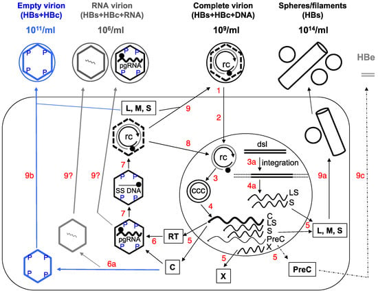
As discussed above, serum HBeAg levels may not strictly correlate with hepatic CCC DNA levels or its transcriptional activity, due to the frequent mutations in the PreC region and core promoter. Therefore, measurement of both serum HBeAg and HBcAg, by using the HBcrAg assay, may not entirely reflect functional CCC DNA in the liver, since CCC DNA defective in HBeAg expression can nevertheless still express HBcAg and the other viral proteins and sustain HBV persistence. Given the aberrant “p22cr” or “HBcrAg” is most likely an artifact, as discussed above, it will be advantageous to measure exclusively serum HBcAg (i.e., empty virions), without the variable contribution of HBeAg. Moreover, as mentioned above, the ratio of empty vs. complete virions in different patients varies widely. Among other factors, this ratio may reflect the efficiency of intrahepatic assembly of empty vs. pgRNA-containing capsids, which, together with the efficiency of reverse transcription and virion assembly and secretion, ultimately determine the ratio of empty vs. complete virions in the blood (Figure 1). Thus, measurement of both complete and empty virions in the blood can also help monitor these events in the liver, which could reflect the changing viral activities, host physiology, and virus-host interactions.

4.1.1。血清空病毒(HBcAg)作为肝癌CCC DNA的标记物
由于空病毒粒子的产生与病毒DNA复制物分离,而是需要HBcAg和HBsAg两者的表达,它们原则上可以作为转录活性CCC DNA的有效生物标志物,在目前用RT抑制剂的抗病毒治疗中,如上所述,可以将血清HBV DNA(即完整病毒体)降低到不可检测的水平,但对CCC DNA水平或其转录活性没有直接影响[78,80]。如上所述,目前的抗病毒治疗中,少数治疗的患者确实经历肝CCC DNA的显着降低,这可能是由于宿主介导的感染细胞或CCC DNA的消除以及治疗诱导的CCC DNA前体的消除(即, RC DNA),从而减少CCC DNA池的补充[78,81,82,83]。在一项试验性研究中,评估血清空病毒颗粒作为肝癌CCC DNA替代生物标志物的潜力,我们使用相对不敏感的蛋白质印迹法(检测限为约50ng / mL)测定空白病毒粒子的血清水平,连同病毒DNA(完整病毒体)和HBsAg水平,在一小组接受RT抑制剂替诺福韦治疗的患者中[9]。通过测量血清HBcAg水平可以方便地监测血清空病毒体的水平,因为完整病毒粒子中HBcAg的量有助于小于总血清HBcAg的1%(即来自完整的和空的病毒粒子)。
在替诺福韦治疗前,血清空白病毒以高达1011 / mL的水平存在,与含RC DNA的病毒体相比超过50至100,000倍[9],这与我们以前在HBV感染黑猩猩和HBV复制肝癌细胞的上清液[8]。在抗病毒治疗之后,如预期的那样,完全病毒体的分泌在几乎所有患者中显着降低(约107 / mL或更多)。然而,即使经过多年的有效的HBV DNA抑制,空肠病毒的分泌在大多数情况下也没有减少。这当然是完全一样预测的,因为DNA合成是分泌完整病毒粒子所必需的,但对于空的病毒粒子分泌是完全无用的,除非肝CCC DNA被消除或稳定沉默,否则它将继续有增无减。事实上,在少数治疗患者中观察到血清空病毒体的显着减少,甚至可能完全丧失,包括那些获得HBsAg损失的患者。结果表明,这些患者可能确实在治疗后肝脏CCC DNA水平(或CCC DNA转录活性)显着减少或减少,尽管相应的肝组织的不可用性不幸地排除了这些患者肝脏CCC DNA的直接测量。将需要具有较大样本量和更敏感的HBcAg测定形式的未来研究以及相应的肝CCC DNA水平的直接测量,以进一步评估血清空病毒粒子作为肝CCC DNA的替代标志物的有用性并确定潜在诊断和血清空病毒粒子的预后意义。由于已知HBc序列在某些区域比其他区域更为可变[84],因此需要认真考虑表位以进行基于抗体的检测以检测血清HBcAg(即空的病毒粒子)。
有趣的是,在实现空白病毒粒子分泌之前,在二十年前尝试测量血清HBc。开发了一种酶联免疫吸附测定(ELISA)试剂盒,用HBc CTD特异性抗体测定血清HBcAg,在未经治疗的患者中,血清HBc和HBV DNA的相关性确实显示出良好的相关性,此外,血清HBc在被监测的单个患者中使用RT抑制剂(在这种情况下为拉米夫定)治疗期间,血清HBV DNA的含量远低于[85]。这些结果导致作者推测无DNA病毒粒子的分泌,尽管它们没有提供任何其他证据。同时,开发了一种不同的ELISA试剂盒,用于测量血清样品中所谓的HBcrAg,已被用于许多临床研究,作为试图监测肝脏CCC DNA的推定替代物。
重要的是要指出,文献中存在一些关于“HBcrAg”是什么的混乱。最初,在2002年,报告了ELISA试剂盒,用于检测从血清样品中的病毒体释放的可溶性HBeAg和HBcAg,使用针对HBc和HBe共有序列的抗体[86]。几年之后,所谓的无异常HBeAg和HBcAg相关蛋白被称为存在于无DNA的HBV中,所谓的p22cr(具有比HBe更长的N-末端延伸和缺失HBc的CTD,见上文)来自HBV感染个体的血清样品中的病毒粒子。这种异常蛋白质也被命名为HBcrAg。在文献中,术语“HBcrAg”用于描述HBc和HBe的组合[13,86,87,88];据说是异常的p22cr [47];和所有三个实体的组合,HBc,HBe和p22cr [89,90,91]。如上所述,所谓的p22cr可能不存在,目前的HBcrAg ELISA试剂盒最有可能检测到HBcAg(从病毒体释放)和HBeAg的组合。因此,当HBeAg存在时,HBcrAg试剂盒检测血清空白病毒粒子和HBeAg,但是在没有HBeAg的情况下,它基本上可以检测到空的病毒粒子(再次,来自完整病毒粒子的血清HBcAg的贡献可以忽略不计)。
不出人意料的是,发现血清HBcrAg水平与血清​​HBV DNA和HBsAg水平相关性相对较低,HBeAg( - )期降低,并且比血清HBV DNA慢得多,并且在血清HBV DNA( - ))患者中仍然可检测到核苷逆转录酶抑制剂(NRTI)治疗[13,88,90],我们观察到血清空病毒体[9]。此外,血清HBcrAg水平与肝脏中HBcAg(+)肝细胞的CCC DNA水平和频率以及肝脏疾病活动相关[87,89,92,93]。还发现预测HBV感染患者的肝癌发展和复发,是比血清HBV DNA更好的预测指标[92,94]。还发现血清HBcrAg与免疫抑制治疗后隐匿性乙型肝炎的再激活相关[91]。
如上所述,由于PreC区域和核心启动子的频繁突变,血清HBeAg水平可能不与肝CCC DNA水平或其转录活性严格相关。因此,通过使用HBcrAg测定,血清HBeAg和HBcAg两者的测量可能不完全反映肝脏中的功能性CCC DNA,因为HBeAg表达中的CCC DNA缺陷仍然可以表达HBcAg和其他病毒蛋白并维持HBV持久性。鉴于异常的“p22cr”或“HBcrAg”最有可能是上述讨论的神器,仅测量血清HBcAg(即空的病毒粒子)是有利的,而HBeAg的贡献不大。此外,如上所述,不同患者的空白对完全病毒粒子的比例差别很大。除了其他因素之外,该比例可能反映了空载体与含有pgRNA的衣壳的肝内装配效率,其与反转录和病毒粒子装配和分泌的效率一起最终决定了血液与空白对完全病毒粒子的比例(图1)。因此,血液中完整和空的病毒粒子的测量也可以帮助监测肝脏中的这些事件,这可以反映病毒活动的变化,宿主生理学和病毒 - 宿主相互作用。

Rank: 8Rank: 8

现金
62111 元 
精华
26 
帖子
30441 
注册时间
2009-10-5 
最后登录
2022-12-28 

才高八斗

6
发表于 2017-3-29 21:14 |只看该作者

Rank: 10Rank: 10Rank: 10

现金
14949 元 
精华
帖子
8576 
注册时间
2008-4-12 
最后登录
2024-5-6 
7
发表于 2017-3-30 20:05 |只看该作者
所谓的p22cr可能不存在???
欢迎收看肝胆卫士大型生活服务类节目《乙肝勿扰》,我们的目标是:普度众友,收获幸福。
我是忠肝义胆MP4。忠肝义胆-战友的天地
QQ群搜"忠肝义胆孰能群"加入

Rank: 8Rank: 8

现金
62111 元 
精华
26 
帖子
30441 
注册时间
2009-10-5 
最后登录
2022-12-28 

才高八斗

8
发表于 2017-3-30 20:32 |只看该作者
回复 MP4 的帖子

也怀疑HBV RNA.

Rank: 10Rank: 10Rank: 10

现金
14949 元 
精华
帖子
8576 
注册时间
2008-4-12 
最后登录
2024-5-6 
9
发表于 2017-3-30 20:49 |只看该作者
StephenW 发表于 2017-3-30 20:32
回复 MP4 的帖子

也怀疑HBV RNA.

不可能,那RNAi全废了
欢迎收看肝胆卫士大型生活服务类节目《乙肝勿扰》,我们的目标是:普度众友,收获幸福。
我是忠肝义胆MP4。忠肝义胆-战友的天地
QQ群搜"忠肝义胆孰能群"加入

Rank: 8Rank: 8

现金
62111 元 
精华
26 
帖子
30441 
注册时间
2009-10-5 
最后登录
2022-12-28 

才高八斗

10
发表于 2017-3-30 20:56 |只看该作者
回复 MP4 的帖子

怀疑含RNA颗粒!
‹ 上一主题|下一主题
你需要登录后才可以回帖 登录 | 注册

肝胆相照论坛

GMT+8, 2024-5-18 16:43 , Processed in 0.016251 second(s), 11 queries , Gzip On.

Powered by Discuz! X1.5

© 2001-2010 Comsenz Inc.